The university is mapping eyes to assess the progression of genetic diseases and the effectiveness of trial treatments
GIS and medical research may not be two fields you’d expect to see join forces. But the University of Oxford and the Oxford Eye Hospital are charting new territory: they’re mapping the eyes of human ophthalmology patients to assess the progress of genetic eye diseases that currently have no cure, and to document the effectiveness of new trial treatments.
The mastermind behind this brilliant innovation, Jasleen Kaur Jolly, MSc BSc (Hons) MCOptom, discovered the potential of GIS applications in the analysis of ophthalmic data through a chance encounter with Jonathan Moules, an FME Certified Professional. This led to an important partnership to explore the use of GIS for ophthalmic data.
Jasleen and her team have been mapping both aspects of the back of the eye to measure how some diseases change the cells in the eye and also aspects of patients’ vision in order to understand how they see the world. Using FME to create a vizualisation of that has been helpful in not only allowing them to see changes over time but also in explaining test results to patients.
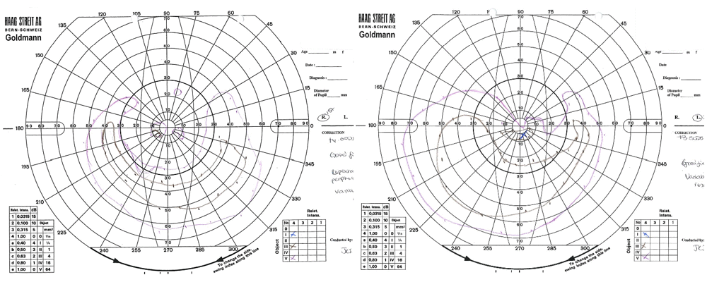
Mapping the visual field of the human eye
Applying GIS technology to choroideremia research
Choroideremia is a genetic disease that causes blindness by the patient’s 30’s, for which there is currently no cure. But the university is hoping to change that with their research related to experimental treatment currently in trial. They are using FME in the quantitative analysis of autofluorescence (AF) scans of the back of the eye. These are images taken with a specialist scanning laser with a wavelength of 488nm and shows up which parts of the retina are alive and which are dying or dead.
Jonathan Moules offered his expertise to the university by creating FME workflows that automatically georeference the scanned raster images of the back of the eye, then extract highlighted areas of the eyes of the 50 patients in the study.
The polygons were then area-on-area overlapped to determine which areas of the eye were most prone to disease. This overlapping method also allowed the university to monitor treatment effect over time. QGIS was then used for creating maps from the shapefiles which had been generated by FME from the overlapped images.
The phase 2 trial for gene therapy has just begun, and will involve a larger cohort of patients to be treated.
Analyzing images for treating Retinitis Pigmentosa
FME has also been used by the university for an experimental treatment for Retinitis Pigmentosa, another genetic disease that results in blindness by early to middle age, and developed a new method of analysis for AF images. FME was used to automatically georeference rasters, taking the resultant images and performing various analyses on them, including creation of terrain profiles, classifications, and axial lines. The results were then used to quantify changes in the disease process, measuring how quickly or slowly the disease changes occurred, and to monitor treatment.
The work with Retinitis Pigmentosa is continuing by improving the quantification methods with a hope to use them in future treatment clinical trials.
The future of FME and ophthalmology
Each disease affects the cells in different ways, and so presents with a different pattern on the pictures taken of the eye. The university are looking to develop methods of image analysis for various diseases with FME while improving the methods they have developed. There has also been interest from others in the field who have seen how FME could help analyze images for their research.
They have found that since ophthalmic imaging contains spatial features, using software that has been designed for spatial data allows them to draw more information from their data and develop innovative ways to analyze it. FME could play a vital role in assessing the success of therapies.
FME has helped them to create protocols for repeatable analysis techniques that can be used across trials, providing comparable results with different treatments. They have also increased their efficiency by automating otherwise laborious parts of the process.
The university’s work has demonstrated that FME can be adapted for ophthalmology, showing that GIS can provide innovative quantitative analyses and allow visualization of various parameters in retinal disease.